About SEMT
The SEMT was founded in 1970 as a forum for the exchange of ideas on techniques and applications in microscopy. It has expanded to become one of the foremost user groups in the country, addressing all aspects of microscopy from instrument design and specimen preparation to digital image acquisition.
We hold an annual meeting with invited speakers and trade stands, facilitating contact, consultation and discussion between all our members, who include instrument manufacturers and microscope users.
We hold an annual meeting with invited speakers and trade stands, facilitating contact, consultation and discussion between all our members, who include instrument manufacturers and microscope users.
Membership
Membership is unrestricted and although mainly London based we have many members from further afield. Membership has benefits for anyone interested in microscopy:
- Newcomers to microscopy
- Experienced microscopists
SEMT promotes co-operation and exchange of ideas as well as the opportunity to keep up-to-date with the latest developments in techniques and equipment.
- Commercial colleagues
SEMT provides an excellent opportunity to advertise to a keenly interested audience and to obtain customer reaction directly.
Officers
Chair
|
Secretary
|
Treasurer
|
Committee members
Ann Dewar
|
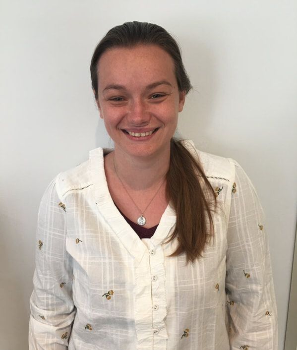
Emily Woodcock |
Jennifer Pearson-Farr
|
|
Jenny is a Senior Laboratory Research Scientist at the Electron Microscopy Science Technology Platform at the Francis Crick Institute. After completing her PhD and postdoctoral position at the University of Southampton, she moved to the Francis Crick to broaden her knowledge in advanced X-ray CT imaging and volume electron microscopy.
|
|
Neil Wilkinson |
|
Paul Gunning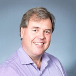
Paul spent 20 years working in colloid science at the BBSRC Institute of Food Research (a.k.a. ‘The Quadram Institute’ from 2018) in Norwich, specialising in electron, light and scanning probe microscopy.
Paul was Head of Surface Analysis (Imaging and Spectroscopy) at Smith+Nephew (global medical devices company) for 19 years, based at Smith+Nephew’s Wound Dressing factory in Hull supporting R&D, Patents and Manufacturing colleagues. The diversity of materials and biological specimen types investigated by Paul’s S+N lab necessitated the use of MSI and several other surface specific analytical techniques, contracted across several external collaborators. Paul joined the University of York in late 2023 and currently uses a wide range of microscopies and spectroscopies including SEM, TEM, SPM, LM, µ-XCT, EDX, µ-XRF, FTIR and Raman for microstructural characterisation and analyses, working across both the Bioscience Technology Facility and the York JEOL Nanocentre to provide internal and external/industrial imaging and spectroscopy services. |
Pedro MachadoSteve Furzeland |
Russell Bailey |
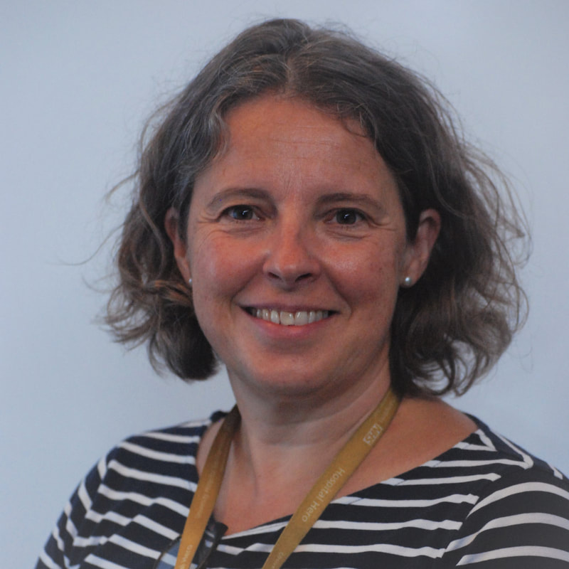
Sarah Hall |